Bio- and Medical Physics
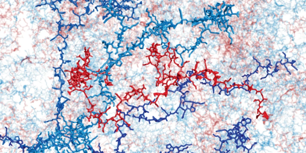
Table of contents
Prof. Christof Aegerter - Disordered and biological soft matter
We study the properties of disordered and heterogeneous systems out of equilibrium. This encompasses light transport in turbid media and photonic glasses, with applications in imaging, structural colours, light harvesting for energy and secure optical communication. A second focus of our activities is the study of the elastic properties of growing biological tissues and their influence on development and pattern formation, e.g. in embryonal development of drosophila, such as ventral furrow formation or dorsal closure. In all these fields our investigations are mainly experimental, developing the tools necessary, e.g. to study forces in tissues on the scale from nN to mN, however we also use computational modeling to guide these experiments.
Ultrafast focusing through turbid media
Imaging structures through turbid media necessitates the creation of a scannable focus after light has passed through the turbid medium. This is possible using spatial modulation of the phase of the incoming light. Using iterative feedback from the transmitted light, creating such a focus is possible using a liquid-crystal spatial light modulator. Here, the optimal phase for each input channel is found sequentially, by determining the phase change of this channel.
This method is however extremely time consuming, as every channel has to be modulated sequentially, such that creating a focus takes several minutes. This effectively makes imaging through dynamic turbid media impossible in this way. We have now created a novel method using a frequency shifted probing beam, which allows for the almost instantaneous determination of the entire complex amplitude of light passing a turbid sample (see Fig. 1). This works because of the frequency shift leads to a beating pattern on the MHz scale, where the phase of the beat corresponds directly to the phase of the input channel. Therefore, a single channel can be optimized within a few microseconds.
Hence, this method allows the creation of a focus on the time scale of 100 ms, optimizing several hundred channels. This can be seen in Fig. 2, where we show focusing through a layer of milk. This shows that using this novel method, imaging through dynamic turbid media becomes feasible.
-

- Zoom (PNG, 484 KB)
- Fig. 1 Left: Setup to determine the intensity as well as the phase of a speckle pattern from light transmitted through a turbid slab (diffuser). The illuminating light is split in an acousto-optical transmission filter (AOTF), creating beams of slightly different frequency. After combining the two beams when passing through the sample, the different frequencies will lead to a beating pattern of the amplitude, whose phase corresponds to the phase difference of the two beams. Right: A speckle pattern determined with the setup in A), where the entire complex amplitude has been determined. The brightness represents the intensity, the colour indicates the phase and the arrows show the whole complex amplitude.
-

- Zoom (PNG, 424 KB)
-
Fig 2 Left: Snapshot of a dynamic speckle pattern created by a sample of milk, where the speckles have a life time of about 0.2 s, as determined by their temporal auto-correlation function.
Right: A focus created by adjusting the spatial phases incurred by passing through the sample using a spatial light modulator. The enhancement of the focus is about 20 times the speckle background and is stable over long times, showing that focusing through dynamic turbid media is possible.
Highlighted Publications:
- A Non-iterative wavefront shaping method focusing light through turbid media,
S. Wang, Master thesis UZH (2024). - Stochastic Resonance in Noisy Optical Resonators Modeled Through Coupled-Mode Theory,
V. Mazzone and C.M. Aegerter, Communications in Nonlinear Science and Numerical Simulation (2024)
Prof. Uwe Schneider - Medical Physics and Radiation Research (Hirslanden)

We are conducting research and development in Medical Physics, Theoretical Biology and Medical Modelling. Our main topics are: Development of radio-biological models, space radiation research, Monte Carlo simulations and dosimetry for radiotherapy and imaging and the development of novel detector systems.
In 2024 our work was focused on two topics. First in Space Ra-
diation Research we tested the use of microdosimeters [1] to determine the biological effect of high-LET radiation at CERN at the HEARTS beamline (see Figure). Second, we developed a novel model for tumor control probability (TCP) modeling in radiotherapy patients. This model allows the transfer of TCP from cohorts of patients to individual patients [2].
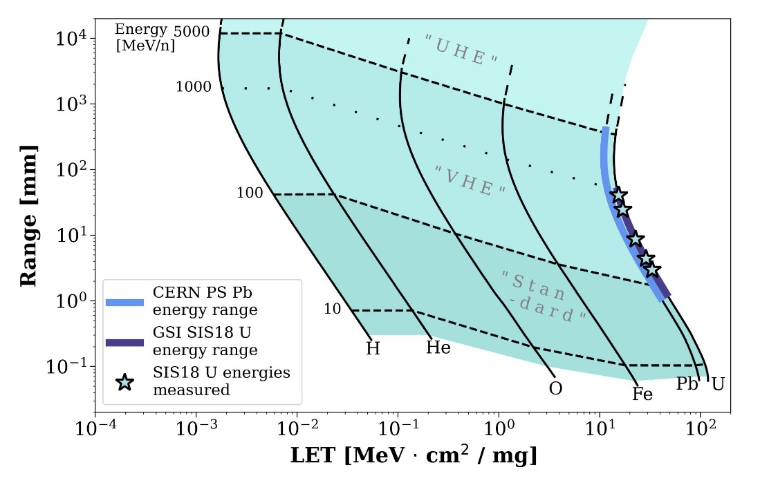
Figure: CERN can offer a “sweet spot” solution to a physical trade-off: high LET (> 30 MeVcm2/mg) combined with a high range (>1 mm) beams at HEARTS (High Energy Accelerators for Radiation effects Testing and Shielding).
Highlighted Publications:
- Microdosimetry of very-high-energy heavy ion beams for electronics testing using silicon-on-insulator detectors
A. Waets et al., IEEE Transactions on Nuclear Science - Realistic closed-form TCP model including cell sensitivity dependence,
KV Diaz Hernandez et al., Submitted to Physics in Medicine and Biology.
Prof. Jan Unkelbach - Medical Physics (University Hospital Zurich)

Radiotherapy is one of the mainstays of cancer treatment and a highly technology-driven field of medicine. In our research group, we contribute to the further development of radiotherapy technology by applying concepts from physics, mathematics, statistics, and machine learning to problems in medical imaging and radiation oncology.
We focus on three areas of research:
1) Radiotherapy treatment planning: We work on mathematical optimization methods to optimally combine x-ray and proton beams, and to optimally distribute radiation dose over multiple treatment days and radiation modalities.
2) Target delineation and outcome prediction: Here, we focus on quantitative modeling of tumor progression and the analysis of medical images such as MRI, CT, and PET, with the goal of precisely defining the region to be irradiated and predicting the patient’s response to treatment (see Figure).
3) Latest radiotherapy technology: We perform research on MR-guided adaptive radiotherapy at the MR-Linac, a combination of MRI scanner and radiotherapy device that allows MR imaging of a patient during treatment.
-

- Zoom (PNG, 92 KB)
- Figure: Modeling of lymphatic metastatic progression of head & neck cancer using statistical machine learning techniques [1]
Highlighted Publications:
- Modelling the lymphatic metastatic progression pathways of OPSCC from multi-institutional datasets.
R. Ludwig et al., Sci Rep. 14(1):15750, 2024.
Prof. Ben Schuler - Molecular Biophysics (Department of Biochemistry)

We study the structure, dynamics, and functions of biomolecules, especially proteins, the nanomachines of life. Towards this goal, we develop and apply single-molecule fluorescence and force spectroscopy, often in close combination with theory and simulations. A particularly important tool is Förster resonance energy transfer (FRET), a spectroscopic nanoscale ruler.
Two highlights were the development of a nanophotonic strategy for enhancing the fluorescence of single molecules [1], and the first measurements of the nanosecond chain dynamics of single-stranded nucleic acids [2].

Figure: Illustration of a nanoantenna with 100 nm gold nanoparticles that enable positioning of a biomolecule for plasmonic fluorescence enhancement. Zoom: Interactions between protein molecules monitored at the nanoscale. Fluorescence time traces (bottom left) show enhanced donor (blue) and acceptor (red) fluorescence during a binding event with unprecedented count rates [1].
Highlighted Publications:
- Single-Molecule FRET at 10 MHz Count Rates,
L. Grabenhorst et al., J. Am. Chem. Soc. 146, 3539–3544 - Nanosecond chain dynamics of single-stranded nucleic acids,
M. Nüesch et al., Nat. Commun. 15, 6010